
Eye Anatomy Explained by Experts
Introduction
Your eyes are among the most complex and remarkable organs in your body, processing over 80% of the information you receive from the world around you. Understanding eye anatomy is fundamental to appreciating how vision works and recognizing the importance of comprehensive eye care. This detailed exploration of eye anatomy will help you understand each component that contributes to your remarkable sense of sight.
At Cannon EyeCare, we believe that patient education about eye anatomy is fundamental to maintaining excellent eye health. This comprehensive guide will walk you through the intricate eye anatomy, explaining how each component works together to create the miracle of sight.
Bottom Line Up Front: Your eye anatomy contains over a dozen specialized structures working in perfect harmony to capture light, focus images, and send visual information to your brain. Understanding these eye anatomy components helps you appreciate why regular comprehensive eye exams at Cannon EyeCare are essential for maintaining lifelong vision health.
How Your Eyes Create Vision: The Process Explained
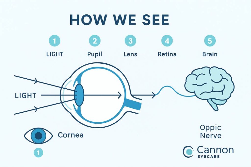
Vision occurs when light enters the eye through the pupil. With help from other important structures in the eye, like the iris and cornea, the appropriate amount of light is directed towards the lens. Just like a lens in a camera sends a message to produce a film, the lens in the eye refracts incoming light onto the retina, where messages are encoded.
The Vision Process in 5 Steps:
- Light Entry: Light enters through the cornea and pupil
- Light Focusing: The cornea and lens bend and focus light rays
- Image Formation: Light creates an image on the retina
- Signal Conversion: Photoreceptor cells convert light into electrical signals
- Brain Processing: The optic nerve carries signals to your brain for interpretation
The eye works much the same as a camera. The shutter of a camera can close or open depending on the amount of light needed to expose the film in the back of the camera. The eye, like the camera shutter, operates in the same way.
The Three Layers of Your Eye Anatomy
Understanding eye anatomy begins with recognizing that the eyeball contains three distinct layers: • The outer layer, formed by the cornea and sclera • The middle layer, holding the primary blood supply for the eye and containing the iris and pupil • The inner layer, comprised of the retina.
Outer Layer (Fibrous Coat)
The protective outer shell that maintains eye shape and provides structural support.
Middle Layer (Vascular Coat)
The nutrient-rich layer contains blood vessels and muscles that control eye function.
Inner Layer (Neural Coat)
The light-sensitive layer where vision begins through specialized photoreceptor cells.
Essential Eye Anatomy Structures and Their Functions
The Cornea: Your Eye’s Clear Window
This protects the inside of your eye like a windshield. Your tear fluid lubricates your corneas. The corneas also do part of the work bending light as it enters your eyes. The cornea is responsible for approximately 65-75% of your eye’s focusing power.
Key Functions:
- Protects inner eye structures from debris and infection
- Focuses incoming light rays onto the retina (provides approximately 65-75% of your eye’s total focusing power)
- Maintains eye pressure through its curved shape
- Filters harmful ultraviolet radiation
Common Issues: Scratches, infections, irregular curvature (astigmatism), and clouding from injury or disease.
The Iris: Your Eye’s Colorful Controller
The iris is the colored portion of your eye. Located behind the cornea and in front of the crystalline lens. This structure separates the anterior and posterior chambers of the eye. The function of the iris is to help regulate the amount of light that enters your eye.
What Determines Eye Color: Eye color is created by the amount and type of pigment in your iris. Multiple genes inherited from each parent determine a person’s eye color.
How It Works:
- In bright light, the iris muscles contract, making the pupil smaller
- In dim light, the iris muscles relax, allowing the pupil to enlarge
- Can change pupil size from 2mm to 8mm in diameter
The Pupil: Your Eye’s Adjustable Opening
The opening in the middle of the iris through which light passes to the back of the eye. Think of your pupil as an automatic aperture that constantly adjusts to optimize vision.
Important Note: Unequal pupil sizes can indicate serious medical conditions and should always be evaluated promptly by an eye care professional.
The Lens: Your Eye’s Focusing System
The lens comprises transparent, flexible tissue and is located directly behind the iris and the pupil. The crystalline lens provides approximately 25-35% of your eye’s focusing power, working together with the cornea to focus light precisely on the retina. Because the lens is flexible and elastic, it can change its curved shape to focus on objects and people nearby or at a distance.
How Accommodation Works:
- Near Vision: Ciliary muscles contract, lens becomes more rounded
- Distance Vision: Ciliary muscles relax, lens flattens
- Age-Related Changes: The lens gradually loses flexibility, leading to presbyopia (difficulty focusing on close objects)
The Retina: Your Eye’s Image Processor
The retina is the light-sensitive tissue that lines the inside surface of the eye, much like wallpaper. Cells in the retina convert incoming light into electrical impulses. These electrical impulses are carried by the optic nerve (which resembles your television cable) to the brain, which finally interprets them as visual images.
Two Types of Photoreceptors:
Rods (approximately 120 million):
- Function in low light conditions (scotopic vision)
- Provide peripheral and night vision
- Detect motion and shapes
- Don’t perceive color
- Outnumber cones by approximately a 20:1 ratio
Cones (approximately 6 million): Cone cells are the second type of light-sensitive cells in the retina of the eye. The human retina contains approximately six million cones; they function best in bright light and are essential for acute vision (receiving a sharp, accurate image). There are three types of cones, each sensitive to different wavelengths: long-wavelength (L-cones/red), medium-wavelength (M-cones/green), and short-wavelength (S-cones/blue).
The Macula and Fovea: Your Central Vision Centers
The macula is the small, sensitive area in the retina’s center that provides clear central vision. The fovea is located in the center of the macula and provides the sharpest detail vision.
Why These Areas Matter:
- Handle detailed tasks like reading and driving
- Contain the highest concentration of cone cells
- Most vulnerable to age-related degeneration
- Critical for legal driving vision requirements
The Optic Nerve: Your Vision Highway
A bundle of approximately 770,000 to 1.7 million nerve fibers carrying visual messages from the retina to the brain. Recent meta-analysis suggests the average number of fibers in each optic nerve is approximately 1.023 million. (To see, we must have light and our eyes must be connected to the brain.) Your brain controls what you see, since it combines images.
Fascinating Facts:
- Contains between 770,000 to 1.7 million individual nerve fibers (average ~1 million)
- Creates a natural “blind spot” where it connects to the retina
- Damage is often irreversible, making early detection crucial
- The main target of glaucoma, a leading cause of blindness
- Shows approximately 3,400 fibers lost per year during normal aging
Supporting Structures: The Unsung Heroes
Sclera: The Protective White Shell
This is the white part of your eye that forms the general shape and structure of your eyeball. The sclera provides attachment points for the six muscles that move your eye and maintains the eye’s spherical shape under varying internal pressures.
Conjunctiva: The Protective Membrane
This clear, thin layer covers the sclera and lines the inside of your eyelids. When this membrane becomes inflamed or infected, it results in conjunctivitis (commonly called “pink eye”).
Vitreous Humor: The Eye’s Gel Filling
The vitreous is the jelly-like substance that fills the inside of the back part of the eye. Over time, the vitreous becomes more liquid and can detach from the back of the eye, creating floaters.
When to Be Concerned:
- Sudden increase in floaters
- Flashing lights in peripheral vision
- Curtain-like shadow in the visual field
- These symptoms may indicate retinal detachment requiring immediate attention
Aqueous Humor: The Eye’s Circulation System
As it circulates, the aqueous fluid flows to the front part of the eye, which is drained by the trabecular meshwork, a sponge-like filtering system where the cornea and iris meet. After draining through the trabecular meshwork, the aqueous fluid then passes through a tiny duct called the canal of Schlemm and is absorbed into the bloodstream.
Why This Matters: Problems with aqueous humor drainage can lead to increased eye pressure and glaucoma, making regular pressure monitoring essential during comprehensive eye exams.
How Your Eyes Work Together: Binocular Vision
Teamwork Benefits:
- Depth Perception: Allows accurate distance judgment
- Peripheral Awareness: Provides approximately a 2a 00-degree field of view
- Backup System: One eye can compensate if the other is compromised
- Enhanced Detail: Both eyes working together improve visual acuity
When Coordination Problems Occur: Issues like amblyopia (“lazy eye”) or strabismus (eye misalignment) can disrupt this teamwork, making early detection and treatment crucial, especially in children.
Age-Related Changes in Eye Anatomy
Natural Aging Process:
20s-30s:
- Peak visual performance
- Maximum lens flexibility
- Optimal tear production
40s-50s:
- Beginning of presbyopia (reading difficulty)
- Slight reduction in pupil size
- Early changes in lens transparency
60s and Beyond:
- Increased risk of cataracts, glaucoma, and macular degeneration
- Reduced contrast sensitivity
- Slower adaptation to lighting changes
When to Seek Professional Care:
Understanding normal aging versus disease symptoms helps you know when to schedule an appointment. Sudden changes in vision, persistent discomfort, or new visual disturbances should always prompt immediate professional evaluation.
Common Eye Anatomy-Related Conditions
Many vision problems stem from variations or changes in normal eye anatomy:
Refractive Errors
- Myopia (Nearsightedness): Light focuses in front of the retina
- Hyperopia (Farsightedness): Light focuses behind the retina
- Astigmatism: Irregular corneal shape causing distorted vision
- Presbyopia: Age-related loss of lens flexibility
Structural Problems
- Cataracts: Clouding of the natural lens
- Glaucoma: Damage to the optic nerve, often from high eye pressure
- Macular Degeneration: Deterioration of central retinal tissue
- Diabetic Retinopathy: Blood vessel damage in the retina
Protective Structure Issues
- Dry Eye Disease: Inadequate or poor-quality tear production
- Conjunctivitis: Inflammation of the eye’s surface membrane
- Corneal Injuries: Scratches or infections of the clear front surface
Latest Research in Eye Anatomy and Function
Recent Scientific Advances (2024-2025)
Current research in eye anatomy continues to reveal new insights about how our visual system works. At Cannon EyeCare, we stay current with the latest developments to provide our patients with cutting-edge care.
Retinal Research Breakthroughs: Retinal ganglion cells (RGCs) are the output neurons of the eye, whose nerve fibers connect through the optic nerve to visual relay centers in the brain. If that connection is interrupted as a result of traumatic injury or ischemic injury, or in neurodegenerative diseases such as glaucoma, there is permanent loss because the optic nerve does not spontaneously regenerate.
Emerging Treatments: Recent studies are exploring regenerative approaches for optic nerve repair, advanced gene therapies for inherited retinal diseases, and stem cell treatments for various eye conditions.
Technology Integration: Modern diagnostic tools like OCT (Optical Coherence Tomography) and advanced retinal imaging allow eye care professionals to detect anatomical changes at the cellular level, often before symptoms appear.
Protecting Your Eye Anatomy: Practical Tips
Understanding eye anatomy empowers you to take better care of your vision. The team at Cannon EyeCare recommends these evidence-based protection strategies:
Daily Protection Strategies
UV Protection:
- Wear quality sunglasses with 100% UV protection
- Consider wraparound styles for maximum coverage
- UV exposure accumulates over time and can damage multiple eye structures
Digital Eye Strain Prevention:
- Follow the 20-20-20 rule: Every 20 minutes, look at something 20 feet away for 20 seconds
- Adjust screen brightness and contrast for comfort
- Consider computer glasses with anti-reflective coatings
Nutritional Support:
- Lutein and Zeaxanthin: Protect macular tissue
- Omega-3 Fatty Acids: Support retinal health and tear production
- Vitamin C and E: Antioxidant protection for various eye structures
- Zinc: Essential for retinal function
When to Schedule Professional Care
Annual Comprehensive Exams: Regular check-ups allow early detection of changes in eye anatomy before they affect vision. Many serious eye conditions have no early symptoms, making professional monitoring essential.
Immediate Attention Needed:
- Sudden vision loss or significant vision changes
- Severe eye pain or persistent discomfort
- New onset of flashing lights or many new floaters
- Curtain-like shadow in peripheral vision
- Any trauma to the eye area
Why Choose Cannon EyeCare for Your Eye Anatomy Health
At Cannon EyeCare, we combine state-of-the-art diagnostic technology with the personal attention and Midwestern hospitality our patients have come to expect. Our comprehensive approach to eye care means we examine every aspect of your eye anatomy to detect problems early and preserve your vision for life.
Our Advanced Eye Anatomy Diagnostic Capabilities:
- High-resolution retinal photography to examine your retinal anatomy
- Optical coherence tomography (OCT) for detailed cross-sectional views of eye structures
- Comprehensive visual field testing to assess optic nerve function
- Advanced dry eye evaluation examining tear film anatomy
- Detailed anterior and posterior segment examination of all eye anatomy components
Convenient Cannon EyeCare Seattle Locations:
- University Village: Modern facility with ample parking and comprehensive eye anatomy evaluation
- Pike Place Market: Downtown convenience for busy professionals seeking expert eye care
Scientific Resources and Citations
This comprehensive eye anatomy guide is based on current medical research and authoritative sources. Here are three key resources that informed this article:
1. Cleveland Clinic – Eye Anatomy and Function (2024)
Source: Cleveland Clinic Medical Center Key Insights: Current understanding of photoreceptor numbers, optic nerve fiber counts, and corneal focusing power percentages. This resource provided updated statistics on rod and cone cell populations and their functional roles in vision. Relevance: Authoritative medical institution providing evidence-based information on eye anatomy structures and their physiological functions.
2. Nature Eye Journal – Optic Nerve Fiber Organization (2024)
Source: Nature Publishing Group – Eye Journal, Volume 38 Key Insights: Recent meta-analysis reveals that human optic nerves contain an average of 1.023 million nerve fibers, with significant individual variation. Research also covers age-related fiber loss patterns and retinotopic organization. Relevance: Peer-reviewed scientific journal providing cutting-edge research on optic nerve anatomy and function, essential for understanding visual pathway integrity.
3. American Academy of Ophthalmology – Optical Properties of the Eye (2024)
Source: American Academy of Ophthalmology Educational Resources Key Insights: Detailed explanation of how the cornea provides approximately 65-75% of the eye’s total focusing power, while the crystalline lens contributes 25-35%. Updated information on accommodation mechanisms and refractive principles. Relevance: Leading professional organization for ophthalmologists, providing authoritative guidelines and educational materials for eye care professionals and patients.
Conclusion
Your eye anatomy represents one of nature’s most sophisticated engineering achievements. Although it is small in size, the eye arguably provides us with the most important of the five senses – vision. Each structure, from the protective cornea to the complex retinal networks, plays a vital role in your ability to see and navigate the world.
Understanding how your eye anatomy works empowers you to make informed decisions about your eye health and recognize when professional care is needed. Regular comprehensive eye exams at Cannon EyeCare don’t just check your vision prescription—they evaluate the health and function of every anatomical structure we’ve discussed.
Don’t wait for vision problems to schedule your next comprehensive eye anatomy evaluation. Contact Cannon EyeCare today to experience the difference that thorough, unhurried attention to your eye anatomy and overall eye health can make.
FAQs
-
The main parts of the eye include the cornea, iris, pupil, lens, retina, macula, optic nerve, and vitreous humor


